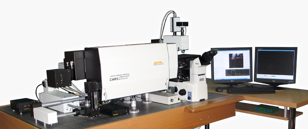
Multifunctionality – confocal 3D scanning laser microscope-spectrometer Confotec® CARS combines in one system:
- CARS scanning microscope
- Raman / luminescent scanning confocal microscope
- Conventional scanning confocal laser microscope
Multi-channel – five channels for simultaneous high-speed measurements:
- F-CARS
- E-CARS & Raman
- reflected laser radiation
- transmitted laser radiation
- luminescent laser radiation
Advantages of CARS method:
High sensitivity: CARS generates more intensive and directed signals in comparison to spontaneous Raman microscopy.
- Anti-Stokes CARS signal has frequency exceeding pumping waves frequencies and is detected in a spectral range free from the stray light of Stokes luminescence.
- CARS signal is registered only in a focus where excitation intensity is the highest. It allows imaging with a high spatial resolution using non-confocal pinholes and also performs 3D layer-by-layer scanning with minimal neighboring layers influence on measuring results.
Spectral resolution of CARS signal is defined only by the width of pumping lasers lines, what simplifies spectral measurements, as detection of CARS signals can be performed without any spectral instrument.
- CARS signal is proportional to the squared molecule concentration, it allows using CARS (along with the selectivity and noninvasivity of the method) for quantitative measurements of chemical substance concentration in a sample.
- Minimal invasive (nondestructive) CARS method for biological samples. Due to the high sensitivity of CARS method molecules in living cells can be detected without fluorescent markers.
Key features
| High spatial resolution: | |
|---|---|
| CARS | XYZ < 0.7 μm |
| Raman | XY < 300 nm Z < 700 nm |
| Wide spectral range: | |
|---|---|
| CARS | 985 – 5000 cm-1 |
| Raman | 75 – 6000 cm-1 |
| High spectral resolution: | |
|---|---|
| CARS | 7 – 8 cm-1 |
| Raman | 0.25 cm-1 |
Simultaneous / multifunctional analysis:
- High-speed imaging by CARS signal (no preliminary additions to the samples).
- Confocal Raman imaging.
- Confocal fluorescence imaging including two-photon (or multi-photon) emission.
- Confocal imaging in the reflected laser radiation.
- High-contrast imaging in the transmitted laser radiation.
- Visualization of a surface profile by means of the 2nd harmonics generation signal.
Three types of CARS measurements:
- F-CARS
- Е-CARS
- P-CARS
Five independent speed channels for simultaneous detection up to four 2D and 3D images:
- F-CARS: CARS signal in forward direction.
- E-CARS & Raman: CARS signal in backward (EPI) direction / Raman signal.
- Reflected: reflected laser radiation signal.
- Transmitted: transmitted laser radiation signal.
- Luminescent: transmitted laser radiation signal.
- Polarization control over the excitation and detection.
- Mono-block laser system for CARS signal excitation.
- Additional laser 633 nm for excitation of a conventional one-photon fluorescence and spontaneous Raman scattering.
- The system is fully PC-controlled: switching of modes of measurement is performed by automatic switching of the components inside the system.
Three scanning modes:
- Laser beam scanning over fixed sample surface with XY scanner.
- Sample transfer by means of XY automated stage relative to the fixed laser beam.
- Multiplexed mode for panorama imaging with a high speed and high spatial resolution: XY scanner + automated stage.
Highly-precise calibration over the wavelengths: better than ±0,002 nm due to an embedded calibration lamp as a source of reference lines for automated operative calibration of the monochromator-spectrograph. Block, rigid framing provides a high temporal and thermal stability.

F-CARS image of polysterene balls of various diameter on frequency 3045 1/cm
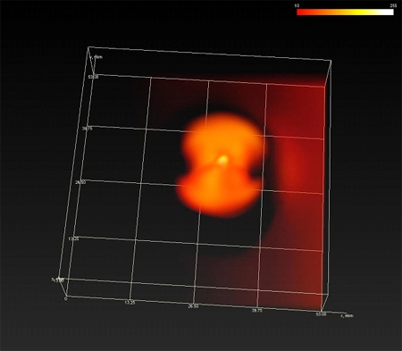
3D CARS image of the liquid crystal 8CB structure on resonant frequency 2236 1/cm

Multimodal CARS/TPEF image of growing cancer cells HeLa: DNA/RNA/Proteins/Lipids colored
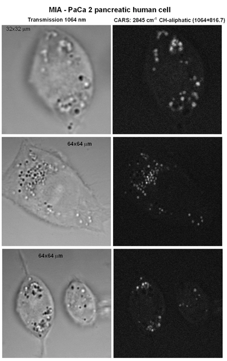
Selective imaging of MIA-PaCa pancreatic human cancer cell for resonance 2845 cm -1 C-H bound (C-H aliphatic stretch). Selective lipids visualization.
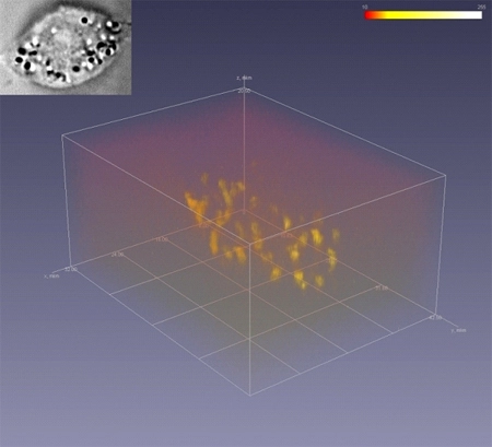
3D image of a cell. References: Dr. A. V. Kachynski, The Institute for Lasers, Photonics, and Bio-photonics, State University of New York at Buffalo.
Intuitive software NanoSP®:
- Online control over the system parameters.
- All-round automation: measuring modes switching by automated switching of the components inside the system; control over the shutters for wavelength selection of the excitation laser; control over the polarization in the excitation/registration channel; selection of a grating, setting of a central wavelength and selection of an output port of the monochromator-spectrograph, adjustment of a pinhole position, etc.
- Confocal 2D and 3D imaging: scanning, accumulation and data storage.
- CARS image, Raman and luminescence spectra.
- Various calibration methods of spectra, also by means of the embedded calibration lamp as a source of reference lines.
- 2D / 3D image of data.
- Image processing:
- Image correction.
- Metric and statistic image processing, free sections.
- Digital filtration, image resize and rotation.
- Spectra processing:
- Background correction (background subtraction).
- Mathematics operations: addition, subtraction, division, multiplication, etc.
- Smoothing with several techniques.
- Searching and definition of spectral lines peaks.
- Scanning modes: Point, XY, XZ, YZ and XYZ.
- Scanning techniques:
- galvanoscanner: 2D high-speed images;
- galvanoscanner + Piezo-Z scanner: 3D high-speed images;
- automated stage: 2D images;
- automated stage + Piezo-Z scanner: 3D images;
- galvanoscanner + automated stage: 2D panorama images;
- galvanoscanner + automated stage + Piezo-Z scanner: 3D panorama images.


Application areas of the 3D scanning laser microscope-spectrometer Confotec® CARS
- Nanobiotechnology: real-time noninvasive analysis of biological samples (cells and components of living cells) with high spatial resolution.
- Micro- and nanotechnology investigations of properties of non-biological microstructures: semiconductors, liquid crystals, polymers, pharmaceutical components, micro- and nanoparticles.


